It began as a scan of the patient's lower jaw in preparation for implants. But what was supposed to be a routine procedure in his Fort Lee, NJ, office turned out to be an event that prosthodontist Scott Ganz, D.M.D., still talks about years later.
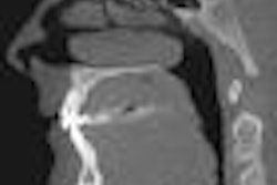
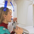
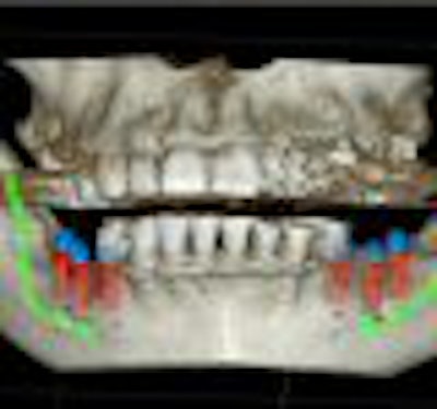
First, the patient was amazed that the scan -- conducted with Dr. Ganz's new cone-beam computed tomography (CBCT) scanner -- was over in a matter of seconds. But more important, the scan revealed a huge cyst in the lower jaw that would obviously need to be removed.
Both the patient and the doctor were pleased that Dr. Ganz had invested in the CBCT scanner, which offers detailed 3D images. The cyst may have gone undetected otherwise, and the system allows Dr. Ganz to monitor the patient's ongoing condition.
"We were alarmed at first," Dr. Ganz said. "Luckily, the cyst turned out to be benign. But because there is a danger of recurrence, we have to scan it periodically. The CBCT device lets us do the work quickly and with relatively low dosages of radiation to the patient."
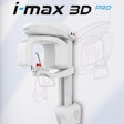
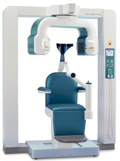
CBCT offers a huge leap forward in dental imaging. For the first time, you can make 3D images in your own office. But any device that carries a price tag of $150,000 to $250,000 needs to be treated as "more than a piece of furniture," in Dr. Ganz's words. In this first of two parts, we examine just exactly what the new technology offers and who should take advantage of it. For anyone planning to do a lot of implants -- and various other specialty procedures -- the investment may pay off quickly. But those dentists more focused on hygiene and routine restorations could wind up paying for razzle dazzle they seldom need.
What is cone-beam CT?
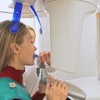
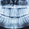
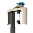
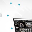
CBCT is a digital x-ray technology used for the head and jaws. It differs from other types of computed tomography (CT) in that multiple diffuse x-ray beams are emitted by the x-ray source. The raw images are then reassembled using a computer algorithm to produce a 3D volumetric image that depicts skeletal, dental, soft tissue, airway, and other internal structures. What's more, it can produce a panoramic scan in as little as 20 seconds.
Yet this new technology hasn't exactly spread like wildfire. In fact, fewer than half of the Kansas dentists who participated in a recent study knew that technology such as cone-beam CT exists. "The most surprising thing," said Anas Athar, D.D.S., an assistant professor of oral radiology at the University of Missouri-Kansas City, "was that, of the 52% who have at least heard about CBCT, few fully understand what it is."
Such ignorance is common because dental schools don't teach enough about the technology, Dr. Ganz. said. And dentists have been reluctant to embrace cone-beam CT because they don't fully appreciate the difference between it and conventional CT. "The problem with regular CT is that you had to send the patient to a radiological center to get the scans," he said. "That required them to go to a huge machine and be exposed to a lot of radiation. Cone-beam CT removes those barriers. You still need education, though. You need to tell students it is a valuable tool."
What cone-beam CT can do for you
Another reason some dentists don't know about cone-beam CT is that it's not for them. If you're a general dentist with few ambitions below the gum line, cone-beam CT could be a waste of your money, said Edwin Parks, D.M.D., M.S., director of dental radiology at the Indiana University School of Dentistry in Indianapolis.
"It's a shotgun when all you need is a fly swatter," Dr. Parks said. "If you have a basic 'drill, fill, and bill' practice, you don't need it." For classic exposures, CBCT may not even work as well traditional intraoral x-rays. The images will be in 3D images but the resolution won't be as sharp as with digital intraoral scanners.
Aside from the big expense (which includes maintenance and possible upgrades to your computer and other equipment), a lot of time may be involved. You and your staff will need training on how to operate the machine, and you may need training on how to interpret the images.
"The single biggest misconception is the time required to learn the new technology," Dr. Ganz said. "A CBCT device is not a turnkey operation. You don't just turn the device on, do a scan, and start reading it. You have to manage the workflow and the flood of information you are going to get."
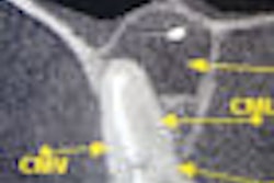
But if you do more complex procedures -- or want to get into them -- then cone-beam could improve your banker's smile as well as those of your patients. "If you are in a practice that does implant placement, it is pretty good," Dr. Parks said. "When you are taking out wisdom teeth, it gives you a good look at the mandibular canal."
CBCT can also help diagnose and plan treatment in periodontics, prosthodontics, oral surgery, orthodontics, TMJ dysfunction, and complex endodontics.
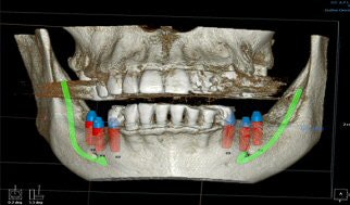
"It allows me to work with more confidence," said Ronald K. Shelley, D.M.D., a general dentist in Glendale, AZ. "I was doing implants and surgery before the cone-beam, but when I got close to a nerve or had to deal with the sinuses, a lot of cases were excluded [because I didn't have] firm evidence of where I was going. Cone-beam gives you 3D information that is really invaluable."
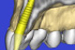
Software such as SimPlant can be used with cone-beam CT to create virtual stents that enable dentists to position implants with precision.
When investigating endodontic lesions, dentists can adjust the angle of the beam to investigate other roots, auxiliary canals, and possible radicular fractures. "With CBCT, you'll do your scans more accurately, without guessing, and with less morbidity to the patient," Dr. Ganz said.
Some orthodonists say CBCT virtually eliminates the need for conventional orthodontic x-rays.
And some dentists who create appliances for sleep apnea say that cone-beam CT helps them visualize the oral-nasal airways. "You need to know where the bone is in three dimensions so you can place it accurately," Dr. Shelley said.
Cone-beam vs. conventional CT
Of course, conventional CT can do most of these tricks as well. So what are the pros and cons of the two technologies?
Perhaps the biggest advantage of cone-beam CT is that you can keep it in your office. Conventional CT machines are too big and too expensive. And having the machine in your office means keeping the patient in your office. First, you won't risk introducing the patients to another practitioner. Second, you'll save them a trip to another facility. Third, you will impress some patients just because you own the latest technology.
And fourth, you can use the detailed images to make a more convincing case for treatment. "You can sell a case that may amount to $10,000 or $20,000 that could never have been marketed any other way," Dr. Ganz said.
But along with the delights of ownership come the headaches. Aside from the time and money involved in ownership, you'll have to take full responsibility for interpreting the images. According to some experts, that means you could be liable for malpractice if you miss a warning sign -- even for a medical problem such as a calcified carotid artery.
Until you are fully trained in reading the output, you may want to send all your images electronically for interpretation. "The images should be interpreted by an oral and maxillofacial radiologist to make sure the volume is thoroughly examined and properly interpreted," said Gail F. Williamson, R.D.H, M.S., a radiology professor at the Indiana University School of Dentistry.
This undermines some of the advantages to owning the machine in the first place. Count on $75 per interpretation -- which brings us back to the hard question of dollars and cents. Just how many images would you need to justify the big ticket of cone-beam CT? For the answers to these questions, look for Part II of this series, coming next week.
