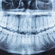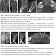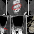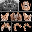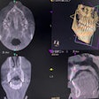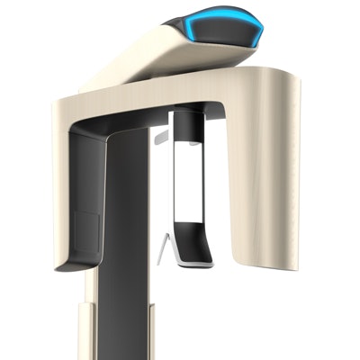
Low-dose cone-beam CT (CBCT) yields statistically similar performance to standard-dose CBCT imaging protocols for CAD/CAM-guided dental implant surgery, according to research published online December 14 in the International Journal of Oral & Maxillofacial Implants.
In a preclinical study conducted on 30 mandibles of pig cadavers, researchers led by Dr. Silvan Unger of the University of Zurich in Switzerland found no significant difference in geometric accuracy between the two techniques.
"Low-dose CBCT imaging protocols providing accurate 3D anatomical information with an improved benefit-risk ratio according to the as low as diagnostically acceptable (ALADA) principle could become a promising option as a primary diagnostic modality as well as for radiologic follow-up," the authors wrote.
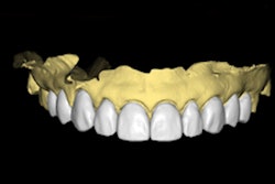
After performing surface scans of the regions of interest, the researchers created a digital diagnostic wax-up and then planned 120 implants, one for each quadrant. Each quadrant was assigned to one of the imaging protocols.
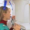
Next, 60 implant surgical guides were manufactured using CAD/CAM technology, and then 60 implants were placed via CBCT guidance in randomized order and following the surgical protocol on an Orthophos SL Unit (Dentsply-Sirona). The authors then assessed the geometric accuracy between the planned and definitive implant position by calculating the apical distances between the central axes and the angle deviation.
| Geometric accuracy of CAD/CAM-guided implants by CBCT imaging protocol | ||
| Standard-dose CBCT | Low-dose CBCT | |
| Implant apical deviation | 0.92 ± 0.55 mm | 0.75 ± 0.63 mm |
| Implant angular deviation | 3.06° ± 2.12° | 2.5° ± 2.12° |
After performing regression analyses, the researchers did not find a significant difference between the two protocols for either of the two measurements.
