Workforce shortages, unhappy patients, and low reimbursements may bring daily dread to dental teams, but the biggest scares may be what happens in the dental chair. In the spirit of Halloween, DrBicuspid reviews its most frightening case reports.
The top case report relates two patients who experienced anaphylaxis, including one who died, after undergoing endodontic treatment. Other petrifying cases include a devastating toothbrush injury, a rare genetic condition that led a baby to have a second mouth, and more.
Without further suspense, here are the 10 most terrifying case reports.
1. Root canal material led to anaphylaxis-related death
Two patients experienced anaphylaxis, one of whom died, after being treated with temporary endodontic material containing polyethylene glycol, according to a case report published in the Journal of Endodontics. This report highlights the possible harm that PEG can cause to some patients and that an allergy to this chemical should be explored in cases of anaphylaxis related to endodontic treatment.
2. Toothbrush injury causes stroke in 2-year-old boy
A 2-year-old boy experienced a blunt cerebrovascular injury to the pharynx that led to an ischemic stroke following a fall while he was brushing his teeth. After treatment and a lengthy hospital stay, the boy remained paralyzed on the lower half of his body on the left side, according to a clinical note published in Pediatrics International.
3. Orthodontic wire travels to a boy’s brain, causing a seizure
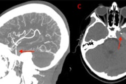
A 12-year-old boy in Texas experienced a seizure after an orthodontic wire from his braces migrated into his temporal lobe.
 (A) and (B) Coronal and sagittal view of the CT angiogram shows the wire penetrating through the skull floor of a 12-year-old boy. (C) Axial view of CT demonstrates the wire in the temporal lobe and the associated intraparenchymal hemorrhage. (D) Axial bone window CT shows the wire entering through the foramen ovale. Image courtesy of Morgan et al. Licensed by CC BY 4.0.
After imaging confirmed the metallic foreign object, which had migrated via the foramen ovale into the temporal lobe, as well as an associated intraparenchymal hemorrhage, the wire was removed without complications. The boy sustained no measurable damage to any structures within or around the foramen ovale, including the carotid artery, which would have been devastating, according to the case report in Radiology Case Reports.
4. Woman develops air in the skull after debridement procedure
A 62-year-old woman developed a life-threatening presence of air within her skull when a dental clinic used an air-polishing device to perform submucosal debridement to treat her peri-implantitis lesion. The case report was published in Clinical and Experimental Dental Research.
Within two seconds of turning on the device, the woman complained of extreme discomfort in her face and head. Computed tomography (CT) scans revealed air in the woman's intracranial space, a phenomenon referred to as pneumocephalus.
 3D reconstruction (a-h) of the emphysema (red) and display of the potential pathways to the intracranial space, i.e., along the inferior orbital fissure and finally via the foramen rotundum (green) and/or via the pterygoid canal (yellow). 3D, three-dimensional. Image courtesy of Bruckmann et al. Licensed by CC BY 4.0.
Due to the air likely being contaminated with oral bacteria, the patient was in danger of developing meningitis. Therefore, she immediately was admitted to the hospital and recovered following antibiotic treatment. This is believed to be the first case of direct development of pneumocephalus from a dental procedure.
5. Root canal sealer found in woman with nerve injury
A 27-year-old woman lost sensation in her chin and lip following root canal treatment on her mandibular molar. Months later, specialists confirmed that extrusion of the root canal sealer caused the patient's permanent anesthesia, according to a case report in Oral Surgery.
 On the left, a dental x-ray shows apical radiolucency, with superimposed radiopaque sealer material extruding at the root tips of endodontically treated tooth #47. On the right, the x-ray shows its presence in socket 47 after extraction. Image courtesy of Mahmood et al. Licensed CC BY 4.0.
Imaging showed that the root canal sealer was widespread within the marrow spaces where endodontic treatment had occurred. The sealer extended to the mandibular canal and perforated the mandible's lingual cortex.
6. Woman loses vision in one eye due to tooth infection-induced cellulitis
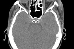
A 54-year-old woman lost vision in one eye after developing orbital cellulitis following a tooth extraction, according to a case report published in Advances in Oral and Maxillofacial Surgery.
 An axial CT scan shows opacification of the woman's left maxillary sinus with marked soft tissue swelling of her left infraorbital. Image courtesy of Ghasemi, et al. Licensed under CC BY 4.0.
CT scans revealed swelling and fluid around the woman's eye and partial opacification of her maxillary sinus and aided in the diagnosis of the rare but deadly bacterial infection odontogenic orbital cellulitis. Surgery treated her infection, but the woman has complete vision loss in her left eye.
7. Teen left with permanent facial scarring after root canal
A 16-year-old girl developed Nicolau syndrome, a rare complication following an intramuscular injection, during a root canal, causing necrosis and leaving her with lasting scars on most of her face. The case report was published in the Journal of Endodontics.
The report was the seventh reported case of an adverse reaction to an intramuscular injection of calcium hydroxide. In 2000, the first clinical case report of this condition involving a 27-year-old man in Belgium was published.
8. Imaging reveals baby girl in SC born with 2nd mouth
Magnetic resonance imaging and CT helped clinicians determine that a mass located on the chin of a newborn girl was a second oral cavity. The cavity contained unerupted teeth and an accessory tongue that moved in sync with her oral tongue, according to a case report published in BMJ Case Reports. The case of diprosopus, an extremely rare congenital disorder in which all or parts of the face are duplicated on the head, is one of about 35 that have been reported since 1900.
9. CPAP use may have led to man’s dental implant prosthesis failure
The use of a CPAP mask and machine may have led to a 71-year-old man's dental prosthesis failure, highlighting this sleep apnea therapy as a possible high-risk factor for implant failure, according to a report published in the Journal of Prosthodontics.
Until the immediate failure of the man's full-arch implant prosthesis, there have been no reported cases of CPAP machine usage as a risk factor for implant failure. After the failure, the man stopped using the CPAP machine for three weeks as instructed by clinicians, and a revision implant surgery was successfully completed.
10. 3-cm swallowed dental needle removed from patient's neck
Clinicians removed a 3-cm needle from a patient who swallowed it, likely during a dental procedure, according to a recent case report published in the Journal of Dental Anesthesia and Pain Medicine. This was believed to be the first reported case of an ingested dental needle causing neck pain.
 A: An x-ray showed a long, thin, and impinged foreign body. B: CT confirmed that the body was metallic and 3 cm in length. C: 3D reconstructed images were used for planning the removal of the needle. Image courtesy of Hassen Mohammed, MD, et al. Licensed under CC BY-NC 4.0.