Improving the look of a patient's papilla can require uncomfortable surgery, but there may be a good alternative, researchers found in a multicenter pilot study Clinical Implant Dentistry and Related Research, October 16 2009.
Researchers from the University of California Los Angeles, the University of Arizona, the University of Washington, the Kois Center, and Hebrew University-Hadassah injected the acid into 14 sites in 11 patients.
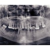
The patients had at least one deficient papilla each in the aesthetic zone, which the researchers photographed before treatment.
The investigators used a 23-gauge needle to inject less than 0.2 mL of an FDA-approved hyaluronic acid gel, 2-3 mm apical to the coronal tip of the papilla in question.
They saw the patients every three weeks and repeated the treatment up to three times, and they tracked the patients from six to 25 months after initial gel application, rephotographing the patients. They used a computer to measure changes in pixels between the before and after pictures. A formula was derived to determine percentage change in the negative space between initial and final examinations.
They found that three implant sites and one site adjacent to a tooth improved 100% between treatment examinations. Seven sites improved from 94% to 97%, three sites improved from 76% to 88%, and one site adjacent to an implant improved 57%.
"Results from this pilot study are encouraging and present evidence that small papillary deficiencies between implants and teeth can be enhanced by injection of a hyaluronic gel," they concluded.