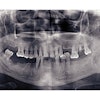
Using digital subtraction radiography (DSR) to assess the outcomes of partial caries removal in deep lesions over a 10-year period, researchers from Federal University of Rio Grande do Sul in Brazil found evidence that this treatment is effective in preventing lesion progression (Oral Surgery, Oral Medicine, Oral Pathology, Oral Radiology, and Endodontology, January 2010, Vol. 109:1, Pp 135-141).
The study prospectively followed 27 patients who underwent partial carious dentin removal. Standardized bitewing radiographs were taken at baseline, with follow-ups at 6-7 months, three years, and 10 years. Tertiary dentin deposition and lesion depth were qualitatively assessed, and radiographic density changes in the radiolucent zone beneath the restoration were quantitatively compared to the control areas using DSR.
A total of 13 teeth were evaluated. In most cases, lesion depth remained unchanged or decreased (12/13) and tertiary dentin formation was observed (10/13) at the 10-year follow-up. Differences between radiolucent zone and control areas at the 6-7-month and 3-year follow-up periods were similar but significantly lower than those at the 10-year assessment. Tertiary dentin deposition was observed in 10 cases at the 10-year follow-up (77%), compared with 5 and 7 cases at the 6-7-month and 3-year examinations, respectively.
"These findings corroborate the statement that, once acid production by the microbial metabolism is controlled, the caries process is controlled as well, regardless of the presence of bacteria in the dental tissue," the researchers wrote. "Our study showed that isolation of microorganisms from the oral environment by cavity sealing is enough to prevent lesion progression."
In addition, they found a high reproducibility rate for quantitative DSR, supporting the reliability of this method for measuring changes in mineral content. Because only substantial mineral loss can be imaged in radiographs, there is a need to develop methods for measuring mineral content with greater accuracy, the researchers noted. In this case, DSR enabled them to detect radiographic density in the radiolucent zone at the 10-year follow-up assessment that was significantly higher than that observed at the 6- to 7-month and 3-year examinations, suggesting mineral gain in this region.
"In this long-term, prospective radiographic study, the sealing of carious dentin was able to: (1) arrest the caries process, demonstrated by unchanged or decreased lesion depth; (2) promote deposition of tertiary dentin; and (3) induce mineral gain in the radiolucent zone," they concluded.